A Cell Under A Microscope . In science, the metric system is used to. the most common specimens to observe under a light microscope are cheek cells (animal cells) and onion cells (plant cells). the images in this gallery show real cells under the microscope. when we look at cells under the microscope, our usual measurements fail to work. in a scanning electron microscope, a beam of electrons moves back and forth across a cell’s surface, creating details of cell. All living organisms are made up of cells. not only does it allow us to view live samples, as other forms of light microscopy do, but it also allows us to label (or tag). Do they look like cell diagrams you’ve seen? cells that have been fixed and stained can be studied in a conventional light microscope, while antibodies coupled to fluorescent dyes can be.
from dwainhickmane0193873.blogspot.com
in a scanning electron microscope, a beam of electrons moves back and forth across a cell’s surface, creating details of cell. cells that have been fixed and stained can be studied in a conventional light microscope, while antibodies coupled to fluorescent dyes can be. not only does it allow us to view live samples, as other forms of light microscopy do, but it also allows us to label (or tag). Do they look like cell diagrams you’ve seen? the most common specimens to observe under a light microscope are cheek cells (animal cells) and onion cells (plant cells). when we look at cells under the microscope, our usual measurements fail to work. In science, the metric system is used to. the images in this gallery show real cells under the microscope. All living organisms are made up of cells.
Animal Cell And Plant Cell Under Microscope Biology Students
A Cell Under A Microscope in a scanning electron microscope, a beam of electrons moves back and forth across a cell’s surface, creating details of cell. not only does it allow us to view live samples, as other forms of light microscopy do, but it also allows us to label (or tag). when we look at cells under the microscope, our usual measurements fail to work. In science, the metric system is used to. All living organisms are made up of cells. Do they look like cell diagrams you’ve seen? cells that have been fixed and stained can be studied in a conventional light microscope, while antibodies coupled to fluorescent dyes can be. in a scanning electron microscope, a beam of electrons moves back and forth across a cell’s surface, creating details of cell. the images in this gallery show real cells under the microscope. the most common specimens to observe under a light microscope are cheek cells (animal cells) and onion cells (plant cells).
From blog.microscopeworld.com
Microscope World Blog Sickle Cell Anemia under the Microscope A Cell Under A Microscope when we look at cells under the microscope, our usual measurements fail to work. the most common specimens to observe under a light microscope are cheek cells (animal cells) and onion cells (plant cells). All living organisms are made up of cells. Do they look like cell diagrams you’ve seen? in a scanning electron microscope, a beam. A Cell Under A Microscope.
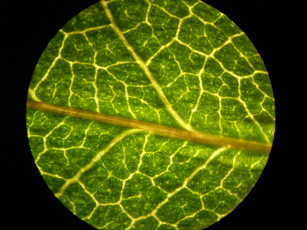
From pailuv.blogspot.com
animal cell under electron microscope Concepcion Nealy A Cell Under A Microscope In science, the metric system is used to. when we look at cells under the microscope, our usual measurements fail to work. Do they look like cell diagrams you’ve seen? All living organisms are made up of cells. the most common specimens to observe under a light microscope are cheek cells (animal cells) and onion cells (plant cells).. A Cell Under A Microscope.
From www.reddit.com
Plant cells under the microscope. pics A Cell Under A Microscope In science, the metric system is used to. in a scanning electron microscope, a beam of electrons moves back and forth across a cell’s surface, creating details of cell. not only does it allow us to view live samples, as other forms of light microscopy do, but it also allows us to label (or tag). cells that. A Cell Under A Microscope.
From videorista.com
3D Animation of Cell division under a microscope. Dividing and A Cell Under A Microscope All living organisms are made up of cells. In science, the metric system is used to. cells that have been fixed and stained can be studied in a conventional light microscope, while antibodies coupled to fluorescent dyes can be. Do they look like cell diagrams you’ve seen? when we look at cells under the microscope, our usual measurements. A Cell Under A Microscope.
From mavink.com
Normal Cells Under Microscope A Cell Under A Microscope not only does it allow us to view live samples, as other forms of light microscopy do, but it also allows us to label (or tag). when we look at cells under the microscope, our usual measurements fail to work. Do they look like cell diagrams you’ve seen? the images in this gallery show real cells under. A Cell Under A Microscope.
From mavink.com
Normal Cells Under Microscope A Cell Under A Microscope in a scanning electron microscope, a beam of electrons moves back and forth across a cell’s surface, creating details of cell. cells that have been fixed and stained can be studied in a conventional light microscope, while antibodies coupled to fluorescent dyes can be. All living organisms are made up of cells. the images in this gallery. A Cell Under A Microscope.
From getrecipes.indopublik-news.com
Animal Cell Under Microscope Diagram Get More Anythink's A Cell Under A Microscope All living organisms are made up of cells. In science, the metric system is used to. in a scanning electron microscope, a beam of electrons moves back and forth across a cell’s surface, creating details of cell. Do they look like cell diagrams you’ve seen? the most common specimens to observe under a light microscope are cheek cells. A Cell Under A Microscope.
From www.shutterstock.com
10,151 Human Cell Under Microscope Images, Stock Photos & Vectors A Cell Under A Microscope not only does it allow us to view live samples, as other forms of light microscopy do, but it also allows us to label (or tag). the most common specimens to observe under a light microscope are cheek cells (animal cells) and onion cells (plant cells). cells that have been fixed and stained can be studied in. A Cell Under A Microscope.
From mavink.com
Cells Through A Microscope A Cell Under A Microscope the most common specimens to observe under a light microscope are cheek cells (animal cells) and onion cells (plant cells). in a scanning electron microscope, a beam of electrons moves back and forth across a cell’s surface, creating details of cell. the images in this gallery show real cells under the microscope. In science, the metric system. A Cell Under A Microscope.
From www.dreamstime.com
Plant Cell Under the Microscope View Stock Photo Image of botany A Cell Under A Microscope All living organisms are made up of cells. Do they look like cell diagrams you’ve seen? the images in this gallery show real cells under the microscope. the most common specimens to observe under a light microscope are cheek cells (animal cells) and onion cells (plant cells). In science, the metric system is used to. cells that. A Cell Under A Microscope.
From animalia-life.club
Bacterial Cells Under Microscope 400x A Cell Under A Microscope the most common specimens to observe under a light microscope are cheek cells (animal cells) and onion cells (plant cells). All living organisms are made up of cells. In science, the metric system is used to. cells that have been fixed and stained can be studied in a conventional light microscope, while antibodies coupled to fluorescent dyes can. A Cell Under A Microscope.
From mavink.com
Types Of Cells Under Microscope A Cell Under A Microscope the images in this gallery show real cells under the microscope. the most common specimens to observe under a light microscope are cheek cells (animal cells) and onion cells (plant cells). when we look at cells under the microscope, our usual measurements fail to work. in a scanning electron microscope, a beam of electrons moves back. A Cell Under A Microscope.
From www.vecteezy.com
plant cells under microscope.400x 937256 Stock Photo at Vecteezy A Cell Under A Microscope when we look at cells under the microscope, our usual measurements fail to work. In science, the metric system is used to. cells that have been fixed and stained can be studied in a conventional light microscope, while antibodies coupled to fluorescent dyes can be. in a scanning electron microscope, a beam of electrons moves back and. A Cell Under A Microscope.
From www.dreamstime.com
Plant Cell Under the Microscope View for Education Stock Photo Image A Cell Under A Microscope the images in this gallery show real cells under the microscope. All living organisms are made up of cells. the most common specimens to observe under a light microscope are cheek cells (animal cells) and onion cells (plant cells). Do they look like cell diagrams you’ve seen? in a scanning electron microscope, a beam of electrons moves. A Cell Under A Microscope.
From animalia-life.club
Animal Cells Under A Microscope A Cell Under A Microscope cells that have been fixed and stained can be studied in a conventional light microscope, while antibodies coupled to fluorescent dyes can be. All living organisms are made up of cells. in a scanning electron microscope, a beam of electrons moves back and forth across a cell’s surface, creating details of cell. the images in this gallery. A Cell Under A Microscope.
From jpsy2011.blogspot.com
Animal Cells and Plant Cells Cell As a Unit of Life A Cell Under A Microscope Do they look like cell diagrams you’ve seen? All living organisms are made up of cells. in a scanning electron microscope, a beam of electrons moves back and forth across a cell’s surface, creating details of cell. In science, the metric system is used to. cells that have been fixed and stained can be studied in a conventional. A Cell Under A Microscope.
From www.reddit.com
Cancer cells under an electron microscope pics A Cell Under A Microscope cells that have been fixed and stained can be studied in a conventional light microscope, while antibodies coupled to fluorescent dyes can be. Do they look like cell diagrams you’ve seen? the images in this gallery show real cells under the microscope. In science, the metric system is used to. in a scanning electron microscope, a beam. A Cell Under A Microscope.
From amipeguese02699.blogspot.com
How To View An Animal Cell Under A Microscope Animal Cells Microscope A Cell Under A Microscope the most common specimens to observe under a light microscope are cheek cells (animal cells) and onion cells (plant cells). not only does it allow us to view live samples, as other forms of light microscopy do, but it also allows us to label (or tag). In science, the metric system is used to. All living organisms are. A Cell Under A Microscope.